Calpas 네비게이터
product
SWIR
Integrated Raman & Hyperspectral Microscopy
제품특징
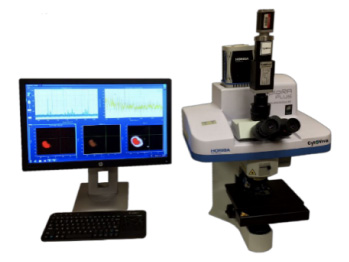
HORIBA Scientific과 CytoViva는 공 촛점 라만 이미징 (Confocal Raman), 하이퍼스펙트럴 이미징 (Hyperspectral) 및 향상된 암시야 이미징 (dark field)을 동일한 현미경 플랫폼에 통합했습니다. 이러한 결합된 양상은 나노 물질 연구에 매우 중요하며, 이 세 가지의 서로 다른 빠른 이미징 방법을 결합하면 나노 스케일에서 관찰대상을 쉽게 관찰, 특성화 및 식별할 수 있습니다. 이 통합 현미경 패키지는 HORIBA Scientific의 입증된 XploRA Plus Raman 현미경을 활용합니다.
이 시스템은 완전 공 촛점 라만 및 PL 이미징을 제공하며 532 nm, 638 nm 또는 785 nm를 포함한 단일 또는 다중 레이저 조명 옵션 중에서 선택할 수 있습니다. 이 통합 이미징 분광계는 전 해상, 범위 및 적용 범위 (격자 600, 1,200, 1,800, 2,400 gr / mm)를 위해 전동 터렛에 장착된 4 개의 격자를 포함합니다. 또한 향상된 감도를 위해 CCD 또는 EMCCD가 포함되어 있습니다. XploRA 수집 및 분석은 사용자에게 친숙한 LabSpec6 소프트웨어 패키지로 수행됩니다. KnowItAll Horiba Raman 라이브러리 검색 소프트웨어는 이 기술과 함께 제공됩니다.
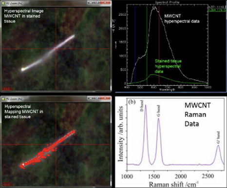
현미경에 CytoViva Enhanced Darkfield 및 Hyperspectral 기술과 HORIBA Raman 기능이 장착되어 있으면 수분 내에 매우 넓은 영역의 하이퍼 스펙트럼 및 darkfield 이미지를 캡처 할 수 있습니다. hyperspectral 이미지는 높은 공간 및 스펙트럼 해상도로 만들어 지며 현미경 접안 렌즈에서 광학적으로 관찰되는 샘플과 매우 흡사합니다. XploRA를 사용하면 동일한 시야에서 선택한 샘플 영역의 라만 스펙트럼을 신속하게 캡처 할 수 있습니다. 단일 샷, 시야를 가로 지르는 선 또는 전체 라만 맵 에서 수행할 수 있습니다. 이는 라만 스펙트럼과 광대역 하이퍼스펙트럴 신호 이미지의 상호 상관을 허용하여 데이터의 정확성을 보장합니다.
단일시료 (탄소 나노 튜브)에 대한 하이퍼 스펙트럼 이미징 분석과 라만분석의 상관교차방식의 분석 사례
Specification
BASE UNIT
| Laser Options Integrated up to 3 internally | 532 nm, 638 nm, 785 nm Other wavelengths and high-power options available on request |
| PC | controlled with Auto Switch option for Raman/white light selection |
| Spectral Resolution | 1.4 cm-1 to 8 cm-1 depending upon grating, laser and CCD selection |
| Spectral Range | 50 cm-1 to 4000 cm-1 depending upon grating, laser selection |
| Spectrograph | Imaging flat field spectrometer for use with larger CCD detectors High throughput with 4 position grating turret |
| Confocal Resolution | Fully confocal, adjustable confocal aperture, 500 nm XY resolution Option for SWIFT™ 10x faster Raman imaging mode |
| Detector | OE. 1024 x 256 pixel TE deep- air cooled -60° C scientific CCD 16 bit. Up to 1.48 MHz readout speed |
MICROSCOPE OPTIONS
Upright Scientific Microscope Includes standard 2 position illuminator with illumination by transmission/reflection. USB image camera
10x and 100x objectives included
Optical Imaging: Standard epi-illumination
Transmitted enhanced darkfield illumination
Objectives Sampling Optics: 50x long working distance, 100x long working distance options
Macro cuvette cell sampler
Fiber-optic probes
Software: OneClick operation as standard
LabSpec Spectroscopy Suite
Options: database, chemometric, imaging, ParticleFinder
ENVIRONMENT - REQUIREMENTS
| Weight | 35 kg - 77 lbs |
| Operating Temperature | 15 - 28° C (optimal 22° C ± 1° C) |
| Voltage | 110/240 VAC, Main supply |
| Dimensions (WxDxH) | 479 x 352 x 661 mm |
| Note | No water cooling or LN2 supply required |